 Menu
Note. Scroll up to view more topics.
Menu
Note. Scroll up to view more topics.
 Home
Home
 Contact Us
 STUDY SKILLS
 Skills of effective study
Making study-notes
Group Discussion
How to concentrate
Developing interest in study
Making preparation for exam
Test Taking Strategies
How to relieve Exam stress?
Time Management for studies
Attempting Computer-based Exam
How to sharpen your brain?
Causes of failure in Exam
Exam-format-wise Preparation
Parents’ Role in Child’s Education
How to improve memory power?
ENGLISH BASICS Noun and its Types
Countable and Uncountable Nouns
Clause
Types of Clauses
Phrase
Types of Phrases
Phrase & Clause - Difference
Verb - Formation & forms of Verbs
Main Verb and Auxiliary Verb
Transitive and Intransitive Verb
Adjective
Comparative and Superlative
Adverb - Use & Formation
Types of Adverb
Pronoun
Types of Pronoun
Prepositional Verb
Coordinating & Subordinating Conjunctions
English Tenses
Active & Passive Voice
Direct & Indirect Narration
TEST PREPARATION IELTS Exam Preparation Guide
CSS Exam, Pakistan
NTS Test (NAT-I, II & GAT)
GRE General Test
MCAT - Medical College Aptitude Test
Entertainment - Funny Jokes Related Topics
Vitamin A – Occurrence & Absorption
Vitamin A – Role & Deficiency Effects
Vitamin D – Occurrence & Absorption
Physiological role of Vitamin D
Effects of Vitamin D Deficiency
Vitamin E – Occurrence & Absorption
Effects of Vitamin E Deficiency
Vitamin C – Changes to it in body
Biochemical role of Vitamin C
Effects of Vitamin C Deficiency
Skills of effective study
Making study-notes
Group Discussion
How to concentrate
Developing interest in study
Making preparation for exam
Test Taking Strategies
How to relieve Exam stress?
Time Management for studies
Attempting Computer-based Exam
How to sharpen your brain?
Causes of failure in Exam
Exam-format-wise Preparation
Parents’ Role in Child’s Education
How to improve memory power?
ENGLISH BASICS Noun and its Types
Countable and Uncountable Nouns
Clause
Types of Clauses
Phrase
Types of Phrases
Phrase & Clause - Difference
Verb - Formation & forms of Verbs
Main Verb and Auxiliary Verb
Transitive and Intransitive Verb
Adjective
Comparative and Superlative
Adverb - Use & Formation
Types of Adverb
Pronoun
Types of Pronoun
Prepositional Verb
Coordinating & Subordinating Conjunctions
English Tenses
Active & Passive Voice
Direct & Indirect Narration
TEST PREPARATION IELTS Exam Preparation Guide
CSS Exam, Pakistan
NTS Test (NAT-I, II & GAT)
GRE General Test
MCAT - Medical College Aptitude Test
Entertainment - Funny Jokes Related Topics
Vitamin A – Occurrence & Absorption
Vitamin A – Role & Deficiency Effects
Vitamin D – Occurrence & Absorption
Physiological role of Vitamin D
Effects of Vitamin D Deficiency
Vitamin E – Occurrence & Absorption
Effects of Vitamin E Deficiency
Vitamin C – Changes to it in body
Biochemical role of Vitamin C
Effects of Vitamin C Deficiency
- Home
- Study Skills
 Skills of effective study Making study-notes Perseverance in study How to concentrate Developing interest in study Group Discussion - Importance Where to study? When to study? Start your study now Making preparation for exam
Preparation according to exam-format
Test Taking Strategies
How to relieve Exam stress?
How to take Computer-based Exam
Making study-notes for exam? Why do students get less marks?
Skills of effective study Making study-notes Perseverance in study How to concentrate Developing interest in study Group Discussion - Importance Where to study? When to study? Start your study now Making preparation for exam
Preparation according to exam-format
Test Taking Strategies
How to relieve Exam stress?
How to take Computer-based Exam
Making study-notes for exam? Why do students get less marks?
- English Basics
Articles
Sentence
Subject, Predicate & Object
Kinds of Sentence (Function-wise)
Kinds of Sentence (Structure-wise)
Phrase & Clause – Comparison
Phrase
Clause
High Frequency words - English
English Tense – Meaning
Present Simple Tense
Present Continuous Tense
Present Perfect Tense
Present Perfect Continuous Tense
Past Simple Tense
Past Continuous Tense
Past Perfect Tense
Past Perfect Continuous Tense
Future Simple Tense
Future Continuous Tense
Future Perfect Tense
Future Perfect Continuous Tense
Active & Passive Voice – Meaning
Passive Voice for Tenses
Passive Voice for Modals
Rules for Imperative Sentences
Sentences which can’t be changed
Direct & Indirect Narration – Meaning
Indirect Speech for Tenses
Rules for Interrogative Sentences
Indirect Speech for Modals
Rules for Exclamatory & Imperatives
Rules for change in Pronoun & Time
- Test Preparation
- Resources
- Contact Us
- Entertainment
- Jobs
- News
Physiological Role of Vitamin A & its Deficiency Effects
The role and deficiency effects of Vitamin A are as follows:
1- Effects on Epithelium
It is needed for the maintenance of healthy epithelium throughout the body. It has been shown to be involved in the formation of mucopolysaccharides. In its absence the normal Epithelium is replaced by a stratified, dry, and keratinized epithelium. This Keratinization results in the following conditions:
- Eye Changes: Here xerophthalmia and keratomalcia occur. Xerophthalamia is characterized by small triangular white patches on the outer and inner sides of the cornea covered by a material resembling white foam; these patches are termed Bitot’s spots. However Bitot’s spots are not specific for vitamin A deficiency and occur in other conditions as well. Keratomalacia occurs usually in the first year of life. The cornea becomes dull and insensitive; the infiltrate increases until finally the cornea necroses ad may appear to melt away within a matter of hours. Keratomalacia is not associated with an inflammatory reaction.
- Changes in Respiratory Tract: Here cilia are lost and respiratory infections take place. This, however, has only been shown in animal and excess of vitamin A does not increase general immunity above normal in human beings.
- Changes in the genitor-urinary tract: The following changes take places in experimental deficiency. Kidney stones may result. This probably caused by an accumulation of stone forming compounds normally present in the Urine around a shed keratinized epithelial cell. In the female, infertility results due to interference with ovulation. In the male rat, atrophy of the germinal epithelium develops. Vitamin A improves reproductive functions in bulls and cows.
- Skin Changes: It becomes dry (xerosis) and shows scaling and small pustules around hair follicles. A popular dry skin eruption called follicular hyperkeratosis, phrynoderma or toad-skin also may be seen.
- Tooth changes: There occurs a defective formation of the tooth enamel and this may lead to abnormalities of dentine.
2- Effects on bones
In the growing animals vitamin A deficiency has been found to result in a retardation of growth accompanied by defects in bone development. The pressure of deformed skull bones on the central Nervous system may produce never degeneration and Paralysis.
3- Role of Vitamin A in Intermediary Metabolism
Vitamin A is involved in the biosynthesis of glucocorticoids. In Vitamin A lack, glucocorticoid synthesis is interfered with; this inhibits the formation of glycogen from such precursors as lactate and glycerol.
An optimum concentration of vitamin A is required for the normal activity of mitochondria and an excess or a deficiency of the vitamin interferes with oxidative phophorylation.
4- Role Vitamin A in Vision in Dim Light
The Isomer 11-cis Vitamin A in the aldehyde form occurs only in Retina. It is essential for rod or night vision. It combines with a protein namely opsin to form a conjugated protein termed “Rhodopsin” or visual purple which is present in rods (in eye ball) and is essential for rod vision. This pigment is very sensitive to light which hydrolyzes it to opsin and all-trans vitamin A aldehyde also called all-trans retinal. The break down of rhodopsin to opsin and all-trans retinal is not as simple as mentioned above. Many intermediate compounds are formed in this process. These include Prelumirhodopsin, lumirhodopsin, metarhodopsin and N-retinylidene opsin. If intense light is thrown into eye all the rhodopsin gets bleached and the person becomes unable to see in the dark. When there is darkness, the synthesis of rhodopsin restarts and after some time the person begins to see in dim light. The regeneration time of rhodopsin depends upon the intensity of light thrown in to the eye and the availability of vitamin A in the body.
The following scheme shows the whole cycle of chemical changes occurring in the rods of the retina.
Physiological Role of Vitamin A
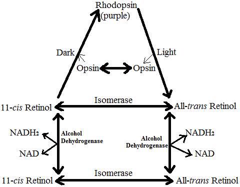
Some loses of Vitamin A occurs in these reaction and is made good by supplies from stores in the body. If vitamin A is not available synthesis of rhodopsin cannot take place and the person becomes unable to see in the dim light (night blindness). It should be noted that not all cases of night blindness are due to lack of Vitamin A
5- Role of Vitamin A in Cone Vision
The cones which are concerned with vision in the bright light as well as with color vision, contain three pigments termed cyanopsin, iodopsin and porphyropsin. All these pigments contain all-trans retinal and the protein opsin which however is different in all these pigments. Cyanopsin, iodopsin and porphyropsin are responsible for seeing three essential colors, i.e. blue, green and red. The mechanism of the breakdown of these pigments almost the same as for rhodopsin.
6- Hypervitaminosis A
Excessive intake of Vitamin A in man produces a number of symptoms especially in infants and young children. When hypervitaminosis A is acute, e.g. after taking a very high dose, headache, nausea, vomiting and drowsiness appear within in a few hours. In the chronic disease caused by daily ingestion of 100,000 units over a prolonged period of the findings are anorexia, dry itchy skin, alopecia, cracking of the lips, and painful areas over the bones. There may be hepatomegaly, splenomegaly, hypothyroidism, leucopenia, anemia, and a bleeding tendency probably caused by hypoprothombinemia. The serum alkaline phosphatase is raised in many cases. These symptoms disappear after the intake of the vitamin is stopped.
Experimentally administration of high doses of vitamin A to pregnant female rats has been shown to result in various congenital defects in the offspring
Storage and Transport of Vitamin A
Vitamin A is stored in the liver mainly as its esters. Kidney and lungs also store vitamin A but to a lesser extent. Vitamin A is carried in blood Plasma by lipoprotein. In protein deficiency the blood level of Vitamin A may be decreased due to the lack of the transporting protein.


Copyright © 2023. STUDYANDEXAM. All Rights Reserved.